|
CLICK TO READ ON IGB'S WEBSITE Living organisms produce a myriad of natural products which can be used in modern medicine and therapeutics. Bacteria and other microbes have become the main source for natural products, including a growing family called ribosomally synthesized and post-translationally modified peptides, or RiPPs. The labs of Douglas Mitchell (MMG), John and Margaret Witt Professor of Chemistry, and Huimin Zhao (CABBI/BSD/GSE/MMG), Steven L. Miller Chair of Chemical and Biomolecular Engineering, at the University of Illinois Urbana-Champaign have been working in tandem to identify and analyze new RiPPs that could be good candidates for drug development and therapeutics. 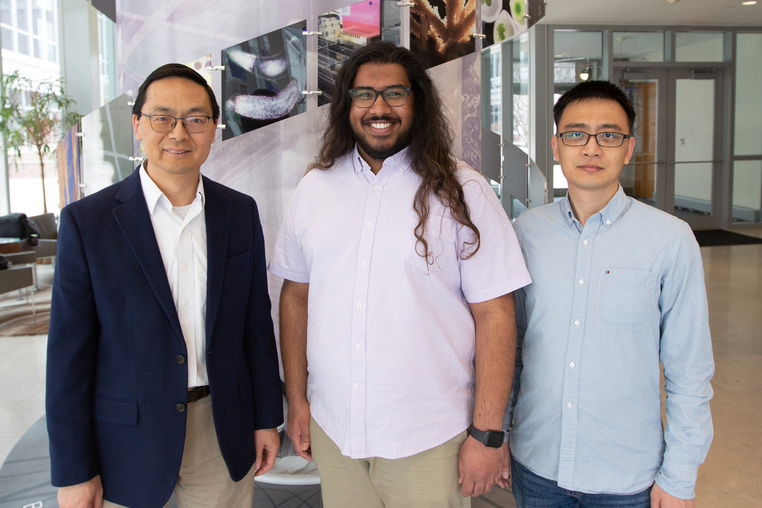 “Compared to other classes of natural products such as alkaloids, terpenes, or polyketides, RiPPs are still underexplored, partly because their biosynthetic gene clusters are quite small and were often overlooked in the past,” said Zhao. “So we decided to develop new technologies to discover novel RiPPs with biological activity.” “Now that we're in the genomics era, we’re realizing just how widespread these groups of natural products are, especially in bacteria,” said Shravan Dommaraju, a PhD candidate in Mitchell’s lab. “We’re basically in this exploration phase, where we know that they're out there, and the goal is to see how many we can find because we don't know what they all do yet.” In a new paper by the Zhao and Mitchell labs, with co-first authors Dommaraju and Hengqian Ren, a postdoctoral researcher in Zhao’s lab, the team reported the discovery of a unique, novel class of RiPPs, which they have named “daptides.” Unlike most peptides which have one positively charged and one negatively charged end, or “terminus,” daptides instead have two positively charged termini. “A textbook would say that a peptide has an amino terminus and a carboxy terminus, but in our case, we found a ribosomal peptide that has two amino termini,” said Ren. “Because there's positive charge at both ends, this gives daptides some interesting bioactivities.” The researchers explained that while this change in termini may seem small, the positive charge of both termini gives daptides the potential to interact with negatively charged objects, such as cell membranes. To test this, the team added the daptides to a dish with red blood cells. They found that the daptides showed hemolytic activity, meaning that they disrupted the membranes of the cells, causing them to rupture. Zhao explained that hemolytic activity is rarely found in the RiPPs the team works with, and that antimicrobial or antifungal activity is much more common. “We were thinking about the structure and what is the evolutionary drive to cause a peptide to lose the negatively charged C terminus and replace it with a positively charged amino group,” Dommaraju said. “From an engineering perspective, if you wanted to make a peptide that can interact with a membrane, you would stick a bunch of positive charges on it. And that's actually what led us to testing this for hemolytic activity because we knew that it had this modification that should allow for that.” Finding new RiPPs is no easy task. First the researchers use bioinformatics to compare and try to identify gene clusters that could produce potential RiPPs. Then they clone the targeted cluster and place it into an organism to be expressed, after which they can check for any natural products produced. Even after obtaining the products, there is still the question of what the products do and how they are produced, which can be tested with bioassays, gene knockouts, and a variety of other tests. But Dommaraju says the labs each play to their expertise when collaborating to streamline the process. “So on a project like this, the Mitchell lab does the bioinformatics and identifies cool gene clusters, and the Zhao lab gets the synthetic biology system up and going to express these peptides and make them,” Dommaraju explained. “So then we are able to tag team our experience and the overlapping stuff that we do to get the project to the finish line.” The researchers say the next steps are to understand the enzyme functions of the daptides, and use bioinformatics analysis to see if there are other combinations of genes associated with daptide production. Directions for further studies in the future include exploring potential therapeutic uses for daptides, and the ecological role that daptide production has for the bacteria that make them. However, both Ren and Dommaraju agreed that their interests in future experiments with RiPPs extends beyond just daptides, as there are still more classes to discover. “We’re interested in using our bioinformatics tool to find as many different natural product classes as we possibly can,” said Ren. “There is such a large frontier right now of undiscovered RiPP classes, and it's exciting to be at the cutting edge, discovering new possibilities. There’s always a chance that the next product you find might be a major therapeutic development!” The paper is published in Nature Communications and can be found at DOI 10.1038/s41467-023-37287-1. The work was funded by National Institute of Allergy and Infectious Diseases (NIAID) and the National Institute of General Medical Sciences (NIGMS).
0 Comments
CLICK TO READ ON IGB'S WEBSITE Similar to how cells within human tissues communicate and function together as a whole, bacteria are also able to communicate with each other through chemical signals, a behavior known as quorum signaling (QS). These chemical signals spread through a biofilm that colonies of bacteria form after they reach a certain density, and are used to help the colonies scavenge food, as well as defend against threats, like antibiotics. 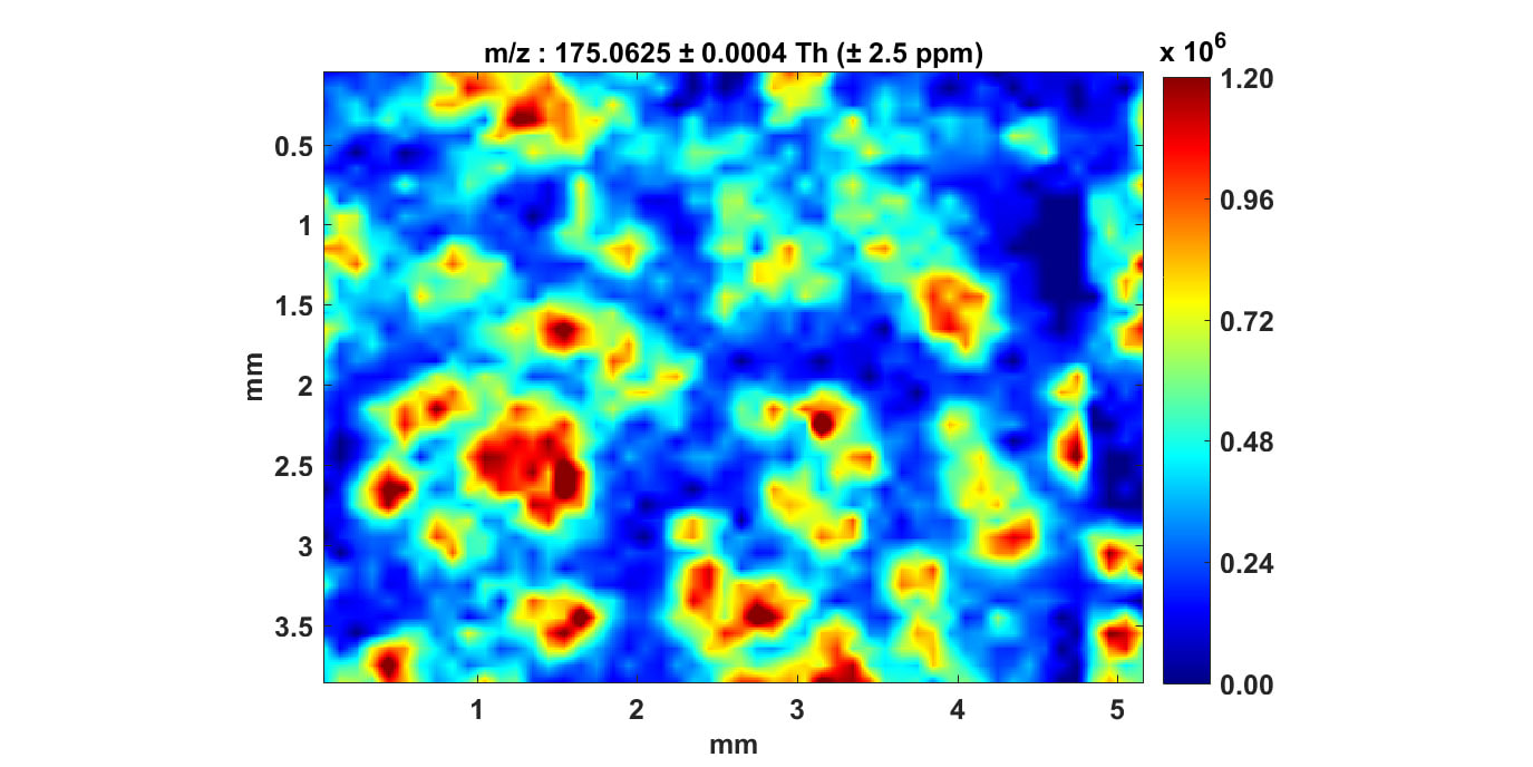 Spatial distribution of Pseudomonas quorum signaling from biofilm sample measured using MS imaging. “QS helps them to build infrastructure around them, like a city,” described Dharmesh Parmar, a postdoctoral researcher in the lab of Jonathan Sweedler (CABBI/BSD/MMG), James R. Eiszner Family Endowed Chair in the department of chemistry. “Biofilms have channels, which allow passage of nutrients, and information in the form of chemical signals. They also allow crosstalk between colonies if there's a threat or the stress in the environment.” Biofilm formation and the subsequent resistance to antibiotics can be especially dangerous for people with a weakened immunity or with diseases like cystic fibrosis (CF), which leads to a stagnant mucus surface inside the lungs that bacteria can more easily attach to. To better understand what surface factors influence or potentially inhibit biofilm formation in the presence of antibiotics, researchers from the Sweedler lab at the University of Illinois Urbana-Champaign, along with collaborators at the University of Notre Dame, measured rate of biofilm formation via QS in a bacteria commonly acquired in hospital infections, Pseudomonas aeruginosa. P. aeruginosa forms biofilms rapidly on a variety of surfaces, which expedites when colonies start communicating using QS, and makes treatment with antibiotics difficult. Additionally, P. aeruginosa can vary in the thickness of the biofilm it produces. The “mucoid” strain produces a thicker biofilm than the non-mucoid strain, and is often linked to infections in patients with CF, a genetic condition which increases mucus viscosity and accumulation in the lungs. In the study, both of these strains were grown on fabricated surfaces that varied in structure, with one being uniform or “unpatterned”, and the other being “patterned” with ridged blocks. Researchers then measured how quickly colonies were able to start communicating with QS while being grown either in the presence of antibiotics or not. QS was detected using mass spectrometry and Raman-imaging, which measured the presence of signaling molecules associated with the behavior. The first thing researchers noticed was that antibiotics slowed growth of a biofilm and production of QS molecules across both strains and structure types. Next, the researchers found that surface type had a large effect on the non-mucoid strain, in that the patterned structure was associated with longer latencies before expression of QS molecules were at their peak. This was not the case for the thicker mucoid strain. “While the impact of the antibiotic slowing biofilm growth didn’t surprise us, the large and differential impact on surface structure was striking,” said Sweedler. “In the non-mucoid strain, surface patterning had a huge impact on the QS signal properties,” Parmar added. “In the case of mucoid, the surface structure had very minimal impact on its metabolic signatures.” The researchers also explored how the distribution of QS signaling molecules differed across different parts of the biofilm when grown on a flat surface and exposed to antibiotics. Samples were taken from the “static biofilm”, where the biofilm attaches to the surface, the “supernatant”, or liquid medium of the culture, and the “pellicle biofilm”, that forms on top of the liquid medium and interacts with the air. Researchers found that the supernatant liquid and pellicle biofilms contained signaling molecules associated with a stress response, while the static biofilm did not contain these molecules. The researchers think this is because the liquid component of the biofilm is what allows bacteria to float along and start new colonies elsewhere, but in the process the bacteria are also exposed to threatening situations, such as presence of antibiotics. By comparing QS behavior during biofilm growth across these different treatments, researchers can better understand how and what kind of molecules this species of bacteria use, and gain new insights on bacterial growth. “P. aeruginosa biofilm is quite challenging to eradicate using currently available antibiotics, and so our goal with this study was to understand what are the factors that govern the growth and stability of these biofilms, and how bacteria escapes these biofilm structures to colonize new locations,” explained Parmar. “The chemically information-rich approaches and analytical techniques we used allowed us to probe these complex molecular events related to biofilm formation across space and time.” Sweedler explained. The team says the next step is to use these optimized analytical techniques to measure QS signals on lung slices from rats, instead of fabricated structures like those used in the current study. Because P. aeruginosa is often associated with infections in the lungs of CF patients, understanding how it forms biofilms in the lungs can help scientists engineer methods to slow or prevent bacteria growth in these patients. Parmar described one potential future application could be to engineer surfaces of medical devices to deter bacterial adhesion and biofilm formation. These findings could also be used to help prevent biofouling, which is when bacteria spoils or degrades biological products and surfaces. The paper is published in ACS Infectious Diseases and can be found at https://doi.org/10.1021/acsinfecdis.2c00519 CLICK TO READ ON IGB'S WEBSITE
James Sharp, one of the industry’s leading authorities in microscopy and former President and CEO of Carl Zeiss Microscopy, has accepted a role as a Distinguished Senior Advisor to the IGB Core Facilities. Sharp obtained his undergraduate degree in electrical engineering before starting his long career at Carl Zeiss Inc. There, he was involved with producing the world’s first electron scanning microscope, as well as installing and calibrating many of the electron and industrial light microscopy instruments currently in use today. Sharp has had numerous management positions in Zeiss microscopy across the world, from Canada, to the United States, to Germany. During these stints he helped coordination acquisition, development, and integration of microscopy businesses and technologies. After this he worked as head of the U.S. based Zeiss Microscopy, where he convinced ZEISS to invest in innovative companies like Cellomics and Atto Instruments, and served as board members for these and other operating companies under ZEISS. In addition to industry connections, Sharp has also networked with many science leaders, including Joe Gray, Lans Taylor, Gary Borsiy, Doug Melton, John Sedat, Jeff Lichtman, and Nobel Laureates Martin Chalfie, Eric Betzig, Guenter Bloebel, Stefan Hell, Harald Hess, and Roger Tsien. Working with these scientists helped Sharp improve light, electron, and x-ray imaging for applications with specific results. Additionally, he was the NA Board Chairman of the German Center for Research and Innovation in New York and is currently a trustee emeritus of the Marine Biological Laboratory. Sharp retired as President and CEO of Carl Zeiss Microscopy, LLC, but still serves as a ZEISS Senior Advisor, where he provides insight into the MicroImaging Market and the commercialization of microscopy. However, his enthusiasm for the sciences has never wavered, and thus he has agreed to bring his talents to Illinois and to the IGB. Sharp’s ties to the institute date back to 2017, when, as President of ZEISS, he started a partnership between the company and the Core Facilities at the IGB. This prestigious partnership allowed the IGB to access state of the art microscopy instruments from ZEISS before others in the field, along with receiving valuable training from ZEISS personnel. Now, as Distinguished Senior Advisor, Sharp will use his intricate knowledge of industry and business surrounding microscopy to help Illinois commercialize system development of microscopy applications, and enhance microscope optics and usage. He will also collaborate with industry representatives and introduce them to Illinois, helping to broaden our reach into the world of industry and beyond. He plans to provide guest lectures for the summer microscopy course, which will hopefully inspire students to consider the wide world of industry as a career path. |
 RSS Feed
